Showing posts with label diatoms. Show all posts

Thursday, May 10, 2012

Things are starting to wind down

The past few days have been kinda slow–but that really awesome kind of slow. The kind of slow where I can take things at an enjoyable pace.

I submitted a second draft of my Master's thesis to my adviser this morning. Once she looks over that, I'll make the necessary changes before I send my thesis to my committee. After I submit my thesis to my committee, I'll be working on my defense presentation. I hope to have my powerpoint almost done before I get back my thesis from my adviser. This would allow me to make minor changes and practice my defense heaps and heaps between submitting my thesis to my committee and giving my defense.

Between there and now, I'll also be doing some bench work trying to get my diatoms in working order for future experiments.

In other news, my supervisor Wendy, who has been advising my work study projects for Clark from blogging to video blogging to Clark videos to photography gave me this sweet travel mug as a parting gift! Such a sweet idea, I know.
#biowithdylan
Even though I won't be moving out until the end of May, I've been eager to start packing a little bit. As a result, my room has been a complete mess of late. It goes through cycles of being very tidy and very messy, at a high rate of turnover. One of my Australian mates had this to say on the Facebook:
Facebook lolz. My room has been a mess because I've been doing some early packing.
While a little breezy, we had some really nice weather today. As such, I decided to take some pictures:

A second mural was painted on Downing St., which is going to be turned into a walking mall. You should check out that link–it talks about the major improvements the Clark campus will experience in the next few months/years. I'm really excited about them all!


Here's another (new) shoot of the first mural, on the corner of Downing and Woodland streets. Atwood is in the background.


This is a shot I've been wanting to take for a while. Below is Estrabrook Hall, on the corner of Woodland and Charlotte streets.

Sunday, May 6, 2012

Bowties and science

While we've had a few nice days recently, it's been pretty rainy over the last few weeks.
Finals are almost over and the students are moving back home. It's kind of a bittersweet time of the semester. The weather and feel of campus brings back memories of just a year ago when I was finishing up my undergraduate career. After being at Clark for 5 year (9 semesters and 2 summers), I'm really going to miss it. I know I'm not looking forward to packing up and moving home myself, but I have plenty to do before then.

I'm defending my Master's thesis on May 22nd, and I hope to be completely done a few days before the end of the month. I have a first draft of my thesis completed, which will probably head over to my thesis committee once my adviser okays it. I've already made corrections from a working (almost complete) draft, which was nearly sufficient for submission anyway. I'm pretty excited about this. Once I submit the draft to the other two members of my committee, I'll focus on finishing my powerpoint presentation.

My powerpoint presentation for my defense will probably be about 45 minutes long. Just thinking about it right now is making me nervous. However, I hope to have a final draft of my presentation completed more than a week before I defend, giving me ample time to practice. This will be the third presentation I've given this semester, and by far the longest, most difficult, and most important. While I'm nervous now and I'll be nervous on my defense day, I just know I'll knock my presentation dead. The closed question period may prove to be much more difficult, but I know I've done more than enough work to complete my Master's.

Between now and the time I move out, I'm almost working on some diatom cultures. I hope to have a large array of diatoms lined up for my professor to work with this summer. Below I discuss part of this process:


And, as a bonus, I made this video in my spare time–how to tie a bowtie!

Thursday, March 15, 2012

Taking 1-step intsead of two.

As of late, there have only been a few things on my mind: the glorious weather, Physiological Ecology of Marine Algae, and 1-step Quantitative PCR.

Spring break ended with spring hitting Worcester with extreme velocity. All of the snow that fell before break has been replaced with highs well into the 60s. There are rumors of temperatures hitting the 70s or 80s next week.

Why yes, I'd love some sunshine while I read.


Aside from the end of this week, with temperatures dropping today and rain expected tomorrow, we've been blessed with sunshine and more warm weather. On Monday I got to spend some time outside while doing some reading during class. While we were given more time than needed to do the reading, I certainly will not complain about the amount of sunrays I was able to soak up while hanging outside. I've beginning to worry about the amount of work I have to do between helping to run two projects in PEMA and finishing up my own research, in addition to preparing presentations, writing my thesis, and giving a defense... all while the weather is getting really nice.

I could try shifting my sleep cycle so that I work a lot at night, sleep in, and enjoy the weather during the afternoon before going back into lab. I highly doubt this is something I'll try. Instead, I'll probably start packing killer lunches and having them on the green in the sunshine.

I have so much to do but I'm still waiting for cultures to grow up and things to be mailed in to retry some of my real-time experiments. I tried running some 1-step QPCR reactions recently since we had the kit on hand. 1-step QPCR makes the cDNA and amplifies it all in one reaction, rather than making the cDNA separately and adding it to the QPCR reaction. While this is definitely easier, saves time, and limits contamination, 1-step QPCR is not as accurate when it comes to estimating the amount of starting template in a sample. As determining the amount of relative template among samples of different test conditions is of the utmost importance to me, it looks like 1-step QPCR won't really fit into my plans any more. However, some initial results suggested my experimental lines of diatoms are behaving as expected (and see previously with normal QPCR), so that's really promising. Once I get a new QPCR kit in and some cells to analyze, my work should be all downhill from here (aside from heaps of lab work).

Watch the video below for a bit more on our beautiful weather and 1-step QPCR.

Sunday, February 19, 2012

Resurrecting old lines of transformed diatoms

I've begun the process of resurrecting old lines of transformed diatoms for future use in experiments in my lab. These diatom cells are from the original agar plates used in the particle bombardment genetic transformations. Currently, I have about 30 discrete lines of diatoms, each with one of my four different experimental plasmid DNA constructs. I'd like to dramatically increase that number if the need arises. The goal is to have a giant pool of diatoms, separated by the type of DNA with which they were transformed.

The overall process is outlined below:

As I stated in the graphic, these lines of diatoms come from the original transformation plates I used to start the discrete lines of diatoms I have now. After using them, I placed them underneath the rack where I grow my diatoms. As this shelf is not a solid plane, limited light did reach these plates. However, the amount of light they did receive was very limited in comparison to the normal growth conditions. This was the only reason why these cells appeared to be alive when I looked at them again recently.

When I was selecting for lines of diatoms back in August/September, I first plated the cells from liquid culture (the original plates I'm discussing now) to be used in the transformation, scraped those cells into liquid culture (much like the diagram above), and then plated the cells again after a recovery period. These cells plated on fresh plates were then left in constant light after they were used to inoculate liquid cultures. After sitting in this light for months on end, they soon faded from their usual brown hue to white. These cells died.

But the original transformation plates, sitting in a dark, cool place, were still brown. Even though they had been sitting on plates without selection (and more importantly without the addition of fresh nutrients), they appeared to still have some life in them.

So I scraped off as many cells from each transformation plate and transferred them into liquid cultures, without any selection. At this point, I had four different test tubes, one for each of my different plasmid constructs transformed into the diatoms. After a week of surprisingly rampant growth, I decided to see if they were still resistant to antibiotics.

Which they were! YEAH SCIENCE!

This past week I have since transferred them to larger liquid cultures to allow the resistant clones to proliferate. I will then plate all of these cells onto multiple selective agar plates, and allow them to grow up before placing them in a cooler, darker place in the culture room.

Until I plate my cultures and select for single colonies (as shown in the last stage of the graphic), I will have "pools" of transformed diatoms for each of my constructs: two different constructs for the nitrate reductase and and nitrite reductase genes. Because of the random insertion of the plasmid DNA into the genomic DNA of the diatom from the particle bombardment, each clone we can separate from the rest of the pool will be distinct from all of the others. This means we have the potential of growing hundreds of different lines of diatoms given the opportunity (or from what was left on the transformation plates).

It will be interesting to see what I do with these lines. If I have time this summer and some money to support me, I may try playing around with different culturing techniques to bolster a manuscript to submit to a journal.

Tuesday, February 7, 2012

New primers and real-time PCR

I've received my new primers and already run a couple different reactions with some results that prove to be promising and others that are frustrating.

Yeah, encouraging :]
The first thing to do with a new set of primers is to run them in a normal PCR reaction to see if the primers amplify the correct length of DNA. While I could do this with my super concentrated, ultra clean plasmid DNA that I used to transform my diatoms (which would serve as the ultimate positive control), I decided to be be bold and try to amplify my complimentary DNA (cDNA) samples. This cDNA was made from mRNA that was extracted from diatom cells exposed to different environmental conditions. Because the mRNA we're looking for contains GFP, anything that is amplified essentially means that our plasmid DNA that we transformed into this line of diatoms is being expressed, which is a great, great thing.

So I ran this PCR reaction with my new primers to amplify my cDNA and this is the gel I got:

There are some pretty convincing bands in that gel which is really encouraging. It appears that all but two of my reactions (6 out of 8) gave us at least some PCR product.

Nicccccce :D
I then ran a real-time PCR reaction called a standard curve, where the source DNA is serially diluted ten fold (I ran 1:1 through 1:10,000 dilutions). For this reaction, I do go ahead and use the plasmid DNA control to give us the cleanest results possible. This reaction allows us to see how efficient the primers are at doubling the amount of DNA product at each PCR cycle.

You can see in this gel (in the first 5 lanes) that a lot of PCR product is being produced. Each reaction hit their saturation point which is why each reaction looks the same even though they had drastically different amounts of starting DNA template. The great this about real-time PCR is that we can see on the computer screen how each reaction was amplified in real time, and see where each reaction it a ceiling amount of DNA. In this reaction I could see that even the 1:10,000 dilution easily hit this ceiling before the 40th (and final) cycle.

With these two encouraging results--the proper amplification using my new primers in a standard PCR reaction and strong amplification in my standard curve using plasmid DNA--I went ahead and tried amplifying my cDNA in a real-time PCR reaction.

Unfortunately, I ran into the same problem I've been having for a few weeks now (see the bottom half of this post).

Sad face real-time PCR :[
Not all of my reactions amplified, and those that did didn't amplify as cleanly as I wanted them to. I ran a gel of my second real-time PCR reaction, which visualizes the two amplified reactions (lanes 3 and 6 starting from the top).

While this is certainly a set back, I have a couple of things I'm going to try. Most importantly, I've ordered a new kit to run the real-time PCR reactions, since the kit I was using was "old." Next, there are a few things I can do to ensure my reactions are as balanced and clean as possible. Finally, I'm going to drop the annealing temperature of my real-time PCR. While I was using the same annealing temperature from my standard PCR, there are two main things that differ between my standard and real-time PCR reactions. First, the salt concentrations are most likely different (although that's a bit annoying to look up, but it's on my to do list), which I found out while screening my diatoms can really screw up a PCR reaction. Second, I used the mysterious "Q-solution" provided by the company Qiagen in their PCR kit when setting up my standard PCR reactions. This may also have significantly changed my standard PCR reaction. While the real-time PCR reaction really should be working with my current set up, it's very possible that my primers are finding it difficult to "seek out" and bind to the sparse cDNA that I want them to amplify.

Monday, January 30, 2012

Extracting RNA from DIATOMZ

In order to measure transcript abundance to collect data for my project, I need to extract RNA from frozen diatom cells. These diatom cells remain "frozen in time" from my experimental assays, which expose lines of diatoms to ammonium. By freezing the cells in liquid nitrogen, I can later extract their RNA and get a good idea of what the transcript abundance was at that time.

To find out more about my experimental assays click on the link up there (which is also here) and then watch my video down below about RNA extractions. These videos together describe some of the most important work for my project!

Sunday, January 29, 2012

Diatom Culling, Revisited

A few weeks ago, I posted about staving off a massive culling of diatoms.

As of right now, my diatoms are still on the mend but I hope to have things all sorted out and lined up by the end of the week. To do this, I'll be checking on my plates and ensuring all of my diatom lines are accounted for before I clean out the culture room, which is in desperate need of being tidied up.

In the video below, I give my account of what it was like to prevent my diatoms from dying. Hopefully I will have been successful in doing so.

Tuesday, January 24, 2012

Designing better RT-PCR primers

Last post I talked about amplifying DNA by real time-PCR, which measures the number of amplified copies at the end of each cycle, giving researchers "real time" numbers of DNA copies. To do this however, you need appropriate primers to get the job done. Not only do they have to be specific enough to work only for the DNA you want amplified, but I'm learning there are other tricks you need to abide by.

Let's back up a little bit though and look at transcription and translation. After all, I'm after the mRNA transcripts that are made in this process. With my genetically engineered construct, protein synthesis starts when transcription factors bind within the cloned 5' untranslated region (UTR) and begin transcription at the promoter, transcribing all of the way through the eGFP open reading frame (ORF) and through the 3' UTR. Now we have an mRNA transcript with part of the 5' and 3' UTR intact at either end of the eGFP coding region. This will serve as the template for translation, which begins at the start codon of the ORF and ends at the stop codon. The 5' and 3' UTR are not translated, hence their UTR moniker.

My project is aiming to measure the amount of mRNA transcript in cells under different environmental conditions. It's not easy to measure mRNA by itself, but it is very easy to measure DNA. Using mRNA as a template, you can make complimentary DNA using the enzyme reverse transcriptase.

Reverse transcriptase starts at the 3' end on an mRNA molecule and transcribes a complimentary strand backwards along the mRNA. However, reverse transcriptase will eventually fall off (represented by the fading orange triangle), so smaller mRNA transcripts work the best.

If I have this mRNA transcript that I want to measure through RT-PCR (after I've converted it into cDNA with reverse transcriptase), I need a primer to amplify the eGFP coding region.

I first made this primer pair to test for the presence of eGFP in my diatom cell lines. It amplifies most of the eGFP coding region by attaching at points just inside of the gene, as you can see below.
Unfortunately, these primers aren't working very well when it comes to applying them to real time-PCR.

My control real time-PCR reactions have worked pretty well for me each and every trial I've run. The control reactions use primers to amplify the endogenous genes we're manipulating in our system, which serve as a good comparison to the experimental reactions.

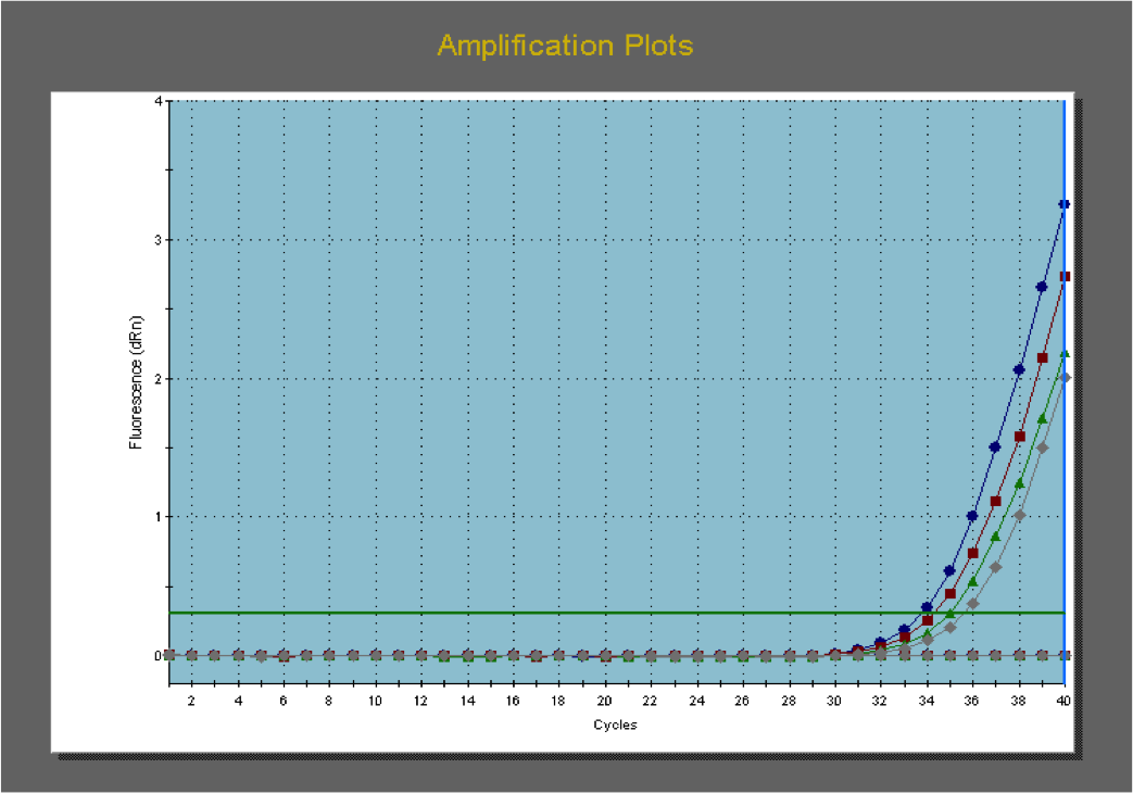
You can see on the graph at right that the primers amplifying endogenous genes work pretty well, developing curves within an appropriate cycle range (the number of cycles until a noticeable amount of product can be measured).

However, when I look at the graph for the transgenic lines using the above primers to amplify eGFP, I get a graph like this on the left. The amplification lines are severely delayed and do not approach the same level of product by the end of the reaction.

In addition to amplification plots, the real-time PCR application on the testing computer also shows graphs that display the melting point of the double stranded DNA molecules. These graphs can be very informative when troubleshooting real time-PCR reactions.

Here is the dissociation curve of the endogenous amplifications:
 Oh boy, it's that curve crisp and clean.

Here is the dissociation curve of the transgenic amplifications:

Yeah, not so much. This curve is extremely messy and non uniform. This provides further evidence that my primers for this reaction might not being working as well as they should be.

After talking to my adviser and seeking some advice online, I've found a couple of parameters to follow to make better primers.

First and foremost (and going back to my bit about reverse transcriptase starting at the 3' end of the mRNA transcripts), my adviser let me in on a secret: I should be using primers that amplify near the 3' end of the transcript since that will be the highest quality region of cDNA as it is transcribed. Using this knowledge, I am working on primers that amplify in the area represented by the orange square, just outside of the 3' UTR. I also tweaked the settings which the primers conform to, based on information I found on other university websites. Yay for Google and other scientists!

The past few days in lab I've been working on these primers and planning out my semester of science ahead of me. Just this week I put together my thesis committee (Justin Thackeray who I had for genetics three years ago, and David Hibbett, my undergraduate adviser) and I've begun re-reading some primary literature and will soon begin reading more broadly and in depth in preparation of writing my thesis. I've also started putting together bits of my paper, which I should have done a while ago.

Anyway, that's all for now, really.

If you've gotten this far, watch my latest YouTubes video regarding this topic matter:

Tuesday, January 10, 2012

Staving off a massive diatom culling

At the end of last semester, chaos tore through my portion of the laboratory when a massive die-off of diatom cultures was a major possibility.

My diatom cultures have been growing in glass test tubes with liquid media, within which they're happy for an upwards of three weeks before they need to be transferred to fresh media. There are a number of factors which make culturing my individual lines of diatoms different from our various stock cultures of diatoms that also grow in the culture room, so it's been a bit of a change for me.

With a few weeks left in the semester, I inoculated two large cultures for my final experimental assay. After about five days, there seemed to be almost no growth, but I figured I'd give them a few more days. After a week I told myself I must have messed up somehow, so I inoculated two more large cultures and I was on my way. At about this time, I made some solid media plates and spread all 40+ of my cultures on individual plates to preserve them while I was away on holiday. (The solid media plates combine the sea water/nutritional supplements I usually grow the diatoms in with agar to solidify the mixture. This is essentially the diatom version of the plates I used to grow my bacteria on when I was cloning DNA.)

But then the second round of large cultures didn't growth either. (Meanwhile my plates still had another 5+ days to show any signs of growth.)

...Yeah... It was at this point I went into a deep panic because my diatoms in my test tubes were dying off and it came to my attention that the materials I was using to supplement my media (i.e. the nutrients to sustain growth) were, well, bad. (This is what happens when I'm not responsible for all of my materials. I am not attacking anyone, but when things matter in the lab I should really do everything myself like make the materials needed for my cultures.) This meant the plates I had made using these materials would not sustain any growth and all of those plates would have to be thrown away.

Now I was upset for multiple reasons: I lost a full day's worth of tedious lab work setting up my first 40 plates and all of my diatom lines were thinking about kicking the bucket.

GREAT.

Additionally, I didn't get in my final experimental assay because my diatoms kept dying in the large cultures due to poor nutritional content.


The end of the semester resulted in frantically making new plates with different materials and pleading with the science gods that they grew while I was away at home.

(Insert a lot of World of Warcraft playing at home)

After a week in the hills of Vermont and New Hampshire, I checked back into lab before heading off to Florida. Thankfully, almost all of my plates showed signs of life which meant I could catch my plane without fretting about my meager diatom cultures.

Attempting to save all of my discrete diatom lines.
After I got back from Florida a week later (roughly two weeks after the second plating), it was time to check on my plates once again.

This time I wasn't as happy with what I saw. Because I was hasty in finishing my plating before I left for home, I did sort of a good-enough-but-crude job. While some of my plates may still be good and support/sustain healthy cultures after they dry off some extra moisture from the initial plating (I sealed each plate to prevent them from drying out), I decided to transfer each culture back to new test tube cultures. I ended up using a combination of plated cultures (two weeks old), old liquid cultures (one month old), and really old liquid cultures (two months old), to sustain almost every line I started with. There are still a few lines I may be able to salvage from my plates
 
A majority of my massive diatom culture collection.
This has been an interesting experience for me and I still have a few things to learn and figure out. The tricky part is that I don't have a lot of spare lab time to tinker around and perfect everything. But because I like everything to be done right, I'll figure them out. First and foremost, I'd like to plate my diatoms so they can subsist for at least three months without me tending to them. While it will be a fair amount of work invested at the beginning of the process, it will save me heaps of time in the long run because I won't have to cater to the high-maintenance demands of my diatoms.

Once I settle back into my hectic life. I hope to consolidate all of my diatom cultures for long-time storage and press onwards to finishing my project for once and for all.

Thursday, November 10, 2011

Extracting RNA from DIE-atoms, among other things

I've been very busy in lab and such, but managed to post a new video onto the YouTubes, which you can see right here:

  
Warning: awesomely awesome intro in this video that's totally worth watching.


It's funny, we had a pretty cold and slightly miserable October that brought us our first snowstorm of the year, but so far November has been very kind to us. Yeah, today it was cold and rainy, but we've had temperatures in the upper 50s and low 60s for most of the month with plenty of sunshine.


While I've enjoyed the beautiful weather on campus, I know all too well it'll get really cold once again and more snow will fall. But until then I'll be sure to appreciate the nice fall weather as long as it lasts.

Sunday, October 23, 2011

Measuring gene activity in changing environmental conditions

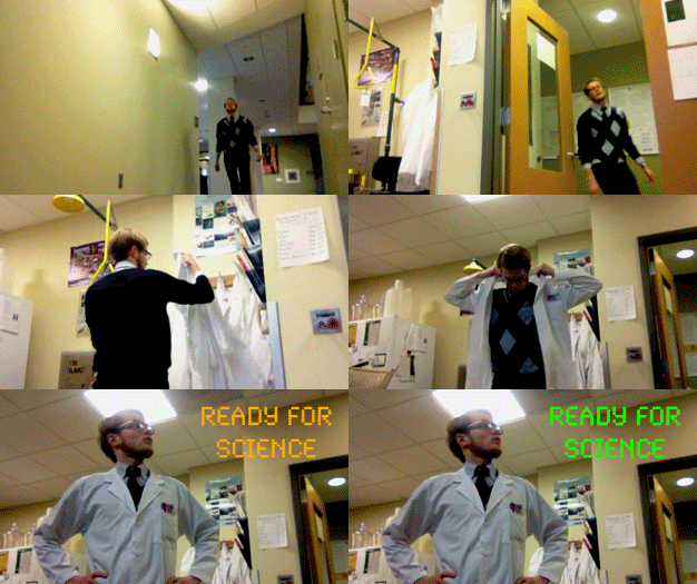
Yeah, so, I'm ready for science.

If I do say so myself, I do look rather badass in this clip. Honestly thought, scientists should be looked up to like super heroes because we're going to be the ones saving the planet (hopefully). More about this topic another day.

Anyway, I've started the phase of my research project that will yield be data. Data for my thesis. Yesssss.

Although my last few videos have been quite long, this one below is short and sweet. How awesome.


My tumblr graphic representing the video >_>


While I still need to plan out all of my assays and assay replicates (to strengthen the power of our collected data), the fact I'm starting this process has me really fired up. For the most part, I'll be running cultures for an hour or so under continuous light (because diatoms like light) under different environmental conditions and taking cell samples periodically for later analysis.

But really, I should be studying for the GRE right now because I take it in less than 3 weeks.

In other brief news:

Last week when I was about to load a gel, I noticed I had a friend. (It's a spider. Click the link!)

In my spare time I put together this Hawk vs. Squirrel video that captures the relationship between the Clark squirrels and the area hawks.

I also updated my Fall @ Clark section, a collection of fall photographs. My most recent additions are at the bottom of the page.

Saturday, October 15, 2011

Diatom Colony Screen

A new lab video!


In this video I discuss designing a colony screen protocol for my transformed lines of diatoms. In one of my recent posts, I posted this picture diagram which oversimplified the process:

While the PCR steps are the same (starting in the top left panel with the diatoms being combined with the PCR mix), I discovered that diatoms grown in liquid culture (i.e. their natural state) needed extra care in preparing them for a colony screen.

Let's step back a bit and quickly talk about colony screens (in case you didn't read my above Tumblr post). A colony screen is a modified PCR reaction*, which is a cyclical amplification of a short sequence of DNA, exponentially copying the targeted DNA strand. (*Even though PCR stands for polymerase chain reaction, PCR is usually said out loud in conversation as PCR reaction.) Often the DNA source for a PCR reaction is a purified, such as plasmid DNA purified by a process to separate plasmid DNA from proteins and genomic DNA.

But colony screens use a colony of cells to supply the DNA for the PCR reaction. By initially lysing the cell by cooking it at a high temperature for a period of time, the cell's DNA is released into the PCR reaction mix and then amplified in the reaction. Colony screens are used to screen cells for the presence of DNA--that is if the targeted sequence of DNA is present, it will be amplified by the reaction. If a reaction gives a positive band on a gel (bottom right of the colony screen chart), then we know that the DNA from the cells in that particular reaction also have the DNA of interest.

This colony screen method should in theory work for other cells, like diatoms. My initial trial successfully amplified GFP from diatom colonies growing on agar plates, but it didn't work for diatom cultures growing in liquid media, a distinction I didn't make at the time.

I soon realized after a failed trial of screening  diatoms solely from liquid cultures that the residual salt water from their growth media was throwing off the delicate chemistry of the PCR reaction. This graphic paired with the above video discusses just this.

Saturday, September 17, 2011

The Genetic Transformation of Diatoms with Inducible Expression Plasmids

Last month my undergraduate/now graduate research project at Clark University reached a new level of awesome with the transformation of diatom cells with plasmids I've been working on for several semesters. It's been a long time coming for sure, with both problems encountered in the lab project and juggling other college courses.

In my blog I've chronicled some steps along the way on my project, and I've talked about:
And now I can talk about the next step of my project: the transformation of diatoms with the plasmids I've created.

Now I am undergoing the process of selecting lines of transformed diatoms for the next step in our project. But we've recently realized that the light intensity in our growth chamber is much brighter than previously published experiments growing transformed cultures of diatoms. Increasing light intensity reduces the amount of chlorophyll made by plants and algae. Why is this important? Two reasons:
  1. Less chlorophyll will reduce the pigmentation of our cells, making them harder to see once we transfer them to liquid culture.
  2. Our gene giving resistance to our antibiotic (that allows us to select for positive transformants) is driven by a chlorophyll-associated protein, which means expression may be reduced in higher light intensities. If chlorophyll expression is reduced by high light levels, then this may reduce the activity of the chlorophyll-associated protein that drives the antibiotic resistance in our diatoms, which means less resistance to the antibiotic in the media. This ultimately means reduced or no growth.
Now, our diatom cultures have been growing okay on their agar plates, but I'm beginning to wonder if they're growing slower in liquid cultures, where they're more likely to absorb more light. To combat this, I'm growing some liquid cultures behind layers of porch screen to reduce light levels penetrating the liquid cultures. In the below picture, you can see the different diatom cultures I have growing right now:

Transformed diatoms grow on agar plates (left foreground) an in liquid media (left background and right).
I've been growing diatoms in 3 mL cultures in little dishes, which you can see in the back left. Groups of 3 dishes sit in a petri dish to allow easier transportation. These dishes however require 3 mL of culture just to cover the entire bottom of the dish, which means the cells are going to be fairly diluted. I also started 1 mL cultures in the test tubes on the right, where porch screen blocks out a lot of the incoming light. Some of the 3 mL cultures also have screen on top of them to block out some of the light.

Aside from trying to grow our cells in liquid culture, I've been looking at them under a fluorescent microscope to try to determine whether our cells are fluorescing due to GFP expression or whether it's residual glow from chlorophyll. UV light from the microscope excites all pigments in the sample, and then I look at the sample through different filters that only allow certain wavelengths of light to show through. However, our current filter set up hasn't allowed a clear delineation between GFP and chlorophyll yet. Most of our pictures look something like this:

...but they usually have a lot more background color in there too.

My next steps will be to either design or order commercial GFP GSPs--gene specific primers for GFP. That way we can run a PCR on some diatom DNA and determine whether they have the GFP gene they are supposed to be transformed with. Because I have a great control (the original plasmid DNA), I can test a whole bunch of diatom colonies at once and be able to select appropriate lines of diatoms faster.

Sunday, September 4, 2011

The start of the semester & culturing transformed diatoms

The backside of Goddard Library, one of my favorite photo spots.
The Fall semester has quickly begun at Clark University. While I was worried that Hurricane Irene was going to make move in day for the sophomores, juniors, and seniors messy, Irene barely inflicted any serious weather upon Worcester (we were lucky). I've been working at the bookstore the past two weeks where it has been very busy, as students have been scrambling to get all of their books on time. As a result, I kind of missed the first week of craziness on campus for better or for worse. But I always love the start of the semester--everyone is always so happy to see each other and be back on campus.

On Friday, we had our annual student activities fair, where almost every social club on campus from CUFS (Clark U Film Society, which shows movies on Tuesdays and Thursdays in our cinema) to ROCU (Radio of Clark U) is represented by student members. This is a great way for incoming first years to discover all of what Clark has to offer for our ever expanding extracurricular groups.

A panorama of the student activities fair, as seen from Main St.
The fair from the window of Tilton Hall, nearly showing the fair in its entirety.
Additionally, I posted a new video on YouTube chronicling the start of the semester, Hurricane Irene in Worcester, and last but not least, my most recent lab work:


Sunday, August 14, 2011

Building inducible expression plasmids (aka Science)

Below is a video I've been trying to publish for a long time coming this summer, but because my summer project only came to a close this week, I've been hesitant to post anything.


In this video, I give an update on my lab work and discuss my overall lab project, which is a great review for myself and my viewers. This is an exciting time for me at Clark University, because I’m beginning the real stages of becoming a graduate student in our accelerated Master’s program. After conducting undergraduate research, I’m using it to support a 5th year of schooling at Clark to finish up my Master’s.

My project concerns the 3’ UTR of the nitrogen-assimilating genes nitrate reductase and nitrite reductase in diatoms (if you haven't read up on my blog before), because we have evidence that along this region these genes are up- and down-regulated by environmental conditions. To test this hypothesis, I’ve created plasmids that drive expression of a reporter gene (GFP) with promoter and terminator regions derived from diatom genes. We’ll compare the activity of GFP with differing 3’ UTR elements to determine whether this region of the nitrogen-assimilating genes is crucial in their regulation.

For the gene nitrate reductase, this is old news. But it is news for my blog that I've completed the plasmids for nitrite reductase, which is really the story of my video.

I'm really excited to be where I am in my project. This week, I hope to genetically transform my diatoms with the plasmids I've created. Oh baby Darwin, this could be a HUGE week for me and my scientific career! I'll be back soon with transformation news.

Thursday, May 26, 2011

More Plasmid Work

I've already talked about how I've made an inducible expression plasmid to test the mRNA stability of nitrate reductase (NR) transcripts in vivo in the diatom Thalassiosira pseudonana. The plasmids run by using a promoter and terminator sequence to run the expression of a reporter gene (we're using GFP). We have a set of NR plasmids, one with a terminal region of NR and one with a terminal region of action (which has nothing to do with nitrogen assimilation). The former plasmid should mimic endogenous activity, whereas the latter plasmid should not mimic the normal conditions in the cell. NR reduces nitrate (NO3-) to nitrite (NO2-), which is then reduced to ammonium (NH4+) by nitrite reductase (NiR). As such, in the scheme of nitrogen assimilation in diatoms, it makes sense to test the regulation of NiR as well.

To do this, I've started work on a set of NiR plasmids. They will also run the inducible expression of GFP, but with their own promoter and terminator regions. Both will have their NiR promoter region, whereas one will have the NiR terminator and one will have the actin terminator region. Both sets of plasmids will be transformed through particle bombardment and then in vivo expression can be measured (through GFP activity).

As it stands, we have a plasmid that contains the NiR promoter & GFP but not the proper terminator. The NiR terminator was cloned into a separate plasmid as part of the process in amplifying out the terminator region of interest. Currently, I'm trying to amplify the terminator out of the plasmid, and in doing so add restriction sites to the plasmid.

The cartoon below represents the series of steps that we have to do in order to manufacture the desired fragment of DNA with restriction sites at the beginning and end of it, which allow us to easily cut out the DNA fragment and place it into a plasmid. Right now, the NiR terminator is represented by the red box. It was amplified out of the entire NiR gene to yield a small piece (between 500bp and 1kb) of the the gene. This piece was at the very end of the open reading frame and extended past the 3' UTR (the terminal region). This was done in a PCR reaction, and the PCR product was inserted into a vector, transformed into bacteria, grown, and then the plasmids were isolated once again the yield the below plasmid.
What I want to do is amplify a smaller portion of the insert out of the plasmid, and make a bunch of copies of it through a PCR reaction. A second set of primers (the green lines) will amplify within the region of the insert, while adding restriction sites. Currently, I've having problems getting this PCR to work because less than half of the primer fits to the DNA of the current insert, while the other half is going to add the restriction site. However, I changed the protocol for the PCR reaction I'm running at the moment. I changed the annealing temperature for the first 10 rounds of my PCR and then I'll bring it back up to what I ran it yesterday (a PCR reaction that did not work, lanes 3-5; right--below right my PCR samples loaded; the faint blue samples are the ladders I used, lanes 1, 2 & 8; the red samples are my PCR reactions that used Coral Load, a special PCR buffer/loading dye combination, lanes 3-7).
...NiR terminator amplification to be continued...

Wednesday, May 25, 2011

Back to Science

While most of my semester was dominated by finishing up my undergraduate courses, I did get a little bit of work done on my "Monitoring in vivo transcription in the marine diatom Thalassiosira pseudonana using eGFP reporter plasmids" project. Most of my work can be summed up through my post on working on the culture side of the transformational protocol (another post here), setting up media for the transformation protocol, and working on my Academic Spree Day poster and a proposal for my summer work on my 5th year Master's project.

I've had really good success on multiple science projects by putting a lot of effort working on a poster or Powerpoint presentation and then writing my term paper for the project. Below is the final draft of my poster (which I hope to hang up outside of the lab later today), which I presented at Academic Spree Day (ASD).

I had a lot of fun at this year's ASD presenting and sharing my work with science (and non-science) professors and classmates. It was particularly satisfying sharing my poster with fellow science (but non-biology) students, and then going over their posters with them. Certainly a little bit of sharing and learning!

This poster then was the basis for updating my project proposal:


This week is my first week after graduating on Sunday. The name of the game for this week is to get back into the flow of things in lab, and hopefully establish a workable rhythm. At the moment, I'm continuing to work on creating inducible expression plasmids for nitrite reductase.

Sunday, March 6, 2011

Spring Break -> no classes -> I can update once again

Classes have been killing me.

I haven't been this busy in a long time. It seems like every week is finals week! I haven't been able to catch a break until this week, which is Spring Break. *catches breath* I don't even know where to start because I haven't done a YouTube video in a month or a real post here in weeks.

Well okay, so this week is spring break which is really nice. How am I spending my week off? Well, my friend Jesse from Pennsylvania is coming up to visit for a few days. I'm wicked stoked he's visiting. He's one of my best friends, and I met him in Australia. Jesse and I were in the same study abroad program in Perth, which I've chronicled in my study abroad blog. Besides his visit, it's catch up time for me. Hopefully I can get a head start on the remainder of the semester, which will be nonstop until May 10th or so. Oh boy. Can you tell I'm excited!?

This will be a long post for sure, so please bear with me.

Directed study

Over the past few weeks, I've been doing culture practice. Because we will be transforming diatoms on plates (to the right), we need to be determine a protocol for growing diatoms on the plates and transferring them to "native" liquid culture. (The top picture are the plates with a poorly drawn circle within which I plated the diatoms [below]. I had to centrifuge down 40mL of culture for each plate, something like a hundred million cells per plate.) I need a little more practice plating the diatoms in the circle outline, and this will ensure maximum efficiency once we do the actual transformation.


In order to plate the cells, I have to count them like I mentioned before. I can then plate a known estimate of cells, and determine what works best. Once I plated the cells and determined how long they took to grow and how few cells I could plate in order to see cultures grow, I needed to transfer them back into liquid culture. This is just like how I grow bacteria on plates and transfer them into liquid culture. But, because the liquid culture for diatoms are much larger than bacteria cultures I use, we need to start the diatoms off in a very small amount, like a few mL. To do this, I took a wire loop and removed a single colony (several hundred cells) and placed it in a 1.5-3.0mL seawater well, on a 6 well plate, which you can see on the middle right in the picture below.

After a few days when I got visible growth, I transferred them into a 5 or 10mL culture, seen in the test tubes. By making larger and larger cultures, we can make sure cells are growing well. If we put our initial cells in a half liter flask, it would take up to a week or two to discern whether we got growth or not. But by growing them in small volumes, we can make sure we're doing okay sooner. (In this picture here you can see my four different diatom culture stages: the plated colonies, bottom left; the test tube 5 and 10mL colonies, top left; the 6 well plate containing 1.5 and 3mL colonies, top right; and the trial transformation plates on the bottom right.)

After break, my professor and I hope to travel down to Rhode Island and transform my diatoms. I can't wait to finally move forward with this project! I'm hoping my post-transformation project will really speed up and I can start collecting data and maybe publish something!

Last but not least,  I was finally accepted into the 5th year biology program, which is really exciting! while I had little doubt I would be accepted, getting the official letter was pretty cool and relaxed me a bit. I got my letter last week, months after other 5th year programs decided whether students could continue their projects or not.

Animal Behavior

A side from reading what I consider to be a lot of papers on different aspects of animal behavior, we're slowly starting to begin our research projects. We have half a semester to collect as much data as possible, write a sophisticated lab report/research paper, and create a lengthy presentation and poster. Something tells me it's going to be an incredible crunch, which is why I'm so eager to get as much studying done as possible this week.

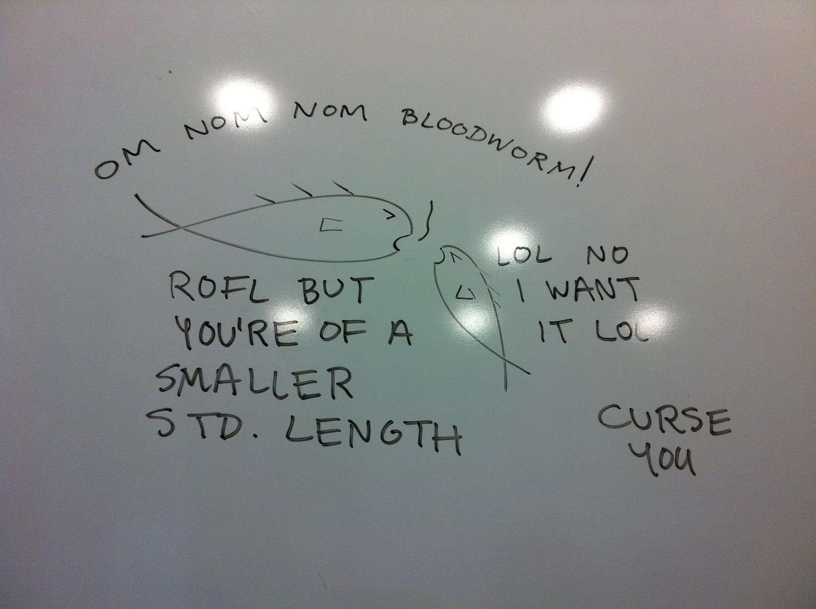
Something I drew on a whiteboard during class...
My research project along with a few friends is to investigate foraging competition among threespine stickleback juveniles, which we call fry. The biggest lab on campus uses threespine stickleback for an array of studies, mostly concerned with evolution and adaptive radiation. But yeah, our project. We're looking into whether body size affects how well stickleback fry can compete for food. In ponds and lakes with limited food sources, competition is likely to be high and we're curious if size is an advantage.


To look at this, we'll be feeding pairs stickleback fry limited amounts of bloodworms, and record their competitions. We do this by pipetting bloodworms into a small tank, and videotaping the fish activity. We can then go back and watch their interactions and analyze it.
However, in order to get them to be competitive, we have to make sure they're hungry... so we don't feed them for half a day before testing.