Showing posts with label diatom transformation. Show all posts

Sunday, February 19, 2012

Resurrecting old lines of transformed diatoms

I've begun the process of resurrecting old lines of transformed diatoms for future use in experiments in my lab. These diatom cells are from the original agar plates used in the particle bombardment genetic transformations. Currently, I have about 30 discrete lines of diatoms, each with one of my four different experimental plasmid DNA constructs. I'd like to dramatically increase that number if the need arises. The goal is to have a giant pool of diatoms, separated by the type of DNA with which they were transformed.

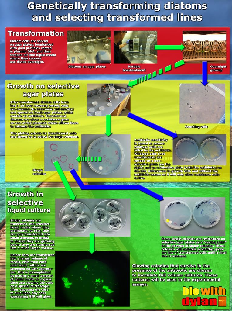
The overall process is outlined below:

As I stated in the graphic, these lines of diatoms come from the original transformation plates I used to start the discrete lines of diatoms I have now. After using them, I placed them underneath the rack where I grow my diatoms. As this shelf is not a solid plane, limited light did reach these plates. However, the amount of light they did receive was very limited in comparison to the normal growth conditions. This was the only reason why these cells appeared to be alive when I looked at them again recently.

When I was selecting for lines of diatoms back in August/September, I first plated the cells from liquid culture (the original plates I'm discussing now) to be used in the transformation, scraped those cells into liquid culture (much like the diagram above), and then plated the cells again after a recovery period. These cells plated on fresh plates were then left in constant light after they were used to inoculate liquid cultures. After sitting in this light for months on end, they soon faded from their usual brown hue to white. These cells died.

But the original transformation plates, sitting in a dark, cool place, were still brown. Even though they had been sitting on plates without selection (and more importantly without the addition of fresh nutrients), they appeared to still have some life in them.

So I scraped off as many cells from each transformation plate and transferred them into liquid cultures, without any selection. At this point, I had four different test tubes, one for each of my different plasmid constructs transformed into the diatoms. After a week of surprisingly rampant growth, I decided to see if they were still resistant to antibiotics.

Which they were! YEAH SCIENCE!

This past week I have since transferred them to larger liquid cultures to allow the resistant clones to proliferate. I will then plate all of these cells onto multiple selective agar plates, and allow them to grow up before placing them in a cooler, darker place in the culture room.

Until I plate my cultures and select for single colonies (as shown in the last stage of the graphic), I will have "pools" of transformed diatoms for each of my constructs: two different constructs for the nitrate reductase and and nitrite reductase genes. Because of the random insertion of the plasmid DNA into the genomic DNA of the diatom from the particle bombardment, each clone we can separate from the rest of the pool will be distinct from all of the others. This means we have the potential of growing hundreds of different lines of diatoms given the opportunity (or from what was left on the transformation plates).

It will be interesting to see what I do with these lines. If I have time this summer and some money to support me, I may try playing around with different culturing techniques to bolster a manuscript to submit to a journal.

Wednesday, November 30, 2011

From GRE to RT-PCR

The view of Lasry the other day on my way into lab.
A few weeks ago I took the GRE as part of my quest to continue graduate school in a Ph.D. program. Standardized testing is such a money scam given all of the fees involved from taking the exam to getting your scores and sending them to your schools. Furthermore, I don't think standardized exams are particularly informative, which is funny because Clark was just in the news because they are no longer requiring SAT scores for undergraduate admissions.

But I digress: I'm not here to complain how ridiculous the whole process of taking the GRE is. I'm just super glad the GRE is behind me and I can focus on things that actually matter. Yet ever since, I've been taking it slow with my workload because I needed a bit of a break. Thanksgiving marked the end of that break though, and now it's time to jump right back into things to finish up a productive semester.

I have a very hopeful list of things I'd like to accomplish by the end of the semester in mid December. But because I'll need to be working a lot more hours at the bookstore to help with the end of semester rush, it's going to be a big squeeze.

We're starting to get books for the spring semester at bookstore, & they're piling up.
This  picture is enhanced with the iPhone app Cat Effects.
Meanwhile in the lab, I have heaps to do. I want to finish up my experimental assays, screen and grow out more diatom cultures for future experiments, prepare a presentation and do some writing, and finally start with some RT-PCR experiments.

Now previously I extracted RNA from my frozen cell samples and used the mRNA present from the cell samples to generate cDNA.

RT-PCR, or real time PCR, will allow me to quantify the cDNA that I have made from my RNA. This is because RT-PCR can measure the number of DNA copies at each copy cycle by measuring the fluorescence of a special dye that hybridizes with the DNA. This is pretty snazzy, but it looks like I'm getting myself into a lot of grunt work.

You see, in order to get precise data from RT-PCR, one usually runs three replicates from one DNA sample and compares the output. But it's not like I only have one DNA sample. I have tons.

This is because I have 8 DNA samples at the minimum per experimental assay that I run in the beginning: I have two different cell lines that I test per assay, which get divided between two different test conditions, from which I take cell samples at multiple timepoints.

For the time being, I'm going to start with time zero and my end time of 60 minutes. This will hopefully give us start and end data that will display the overall trend of my experiments.

But because of positional effects, I need to run more than one experimental assay. I'll probably run three sets of samples through RT-PCR, but I hope to have up to 5 samples completed and ready for RT-PCR by the end of next week.

This is from today while I waited for my next sampling time.
Wait, what's that positional effects you just mentioned a second ago? Oh, yeah, positional effect. Because we transformed our diatoms with ballistics, our DNA was randomly inserted into the diatom's genome. By chance, the DNA we're trying to measure in our experiments might be inserted into a region that is either expressed more or less often, which would skew our data. By gathering data from multiple replicates, we'll get more accurate data regarding gene expression. Cool how science works, huh? We think of the neatest little caveats. Well, I mean, my professor does.

But sometimes I do too!

Like last month or so I came up with a solution as to why our cultures weren't growing so well. And then more recently I figured out how to screen my diatoms for our reporter gene more effectively.

So yes, sometimes I come up with cool things too.

The Jonas Clark building. I can't stop taking pictures from this vantage point! Gah! I love it! 11/18/2011

Tuesday, September 13, 2011

Glowing Green Cells

Just before the weekend, my professor wanted to look under the microscope to see whether our transformed diatom cells were expressing our reporter gene GFP. I've been culturing several different lines of transformed diatoms since the transformation. These cells were co-transformed, which means the resistance plasmid is separate from the GFP plasmid--that is the transformation process shot two different plasmids at the diatom cultures. Therefore we need to select for diatom lines that posses both plasmids: the resistance plasmid and the GFP plasmid.

Below is a poster I made briefly describing each of the three main steps in the process: transformation, growing the diatoms on selective agar plates, and subculturing diatom cells in selective liquid cultures.

The diatoms will only grow if they received a copy of the resistance plasmid. And well, we can tell that the diatoms don't particularly like the antibiotic we put in the growth media:

By performing a simple antibiotic sensitivity test, we can show that diatoms will not survive in the presence of the antibiotic unless they receive the plasmid that confers resistance.

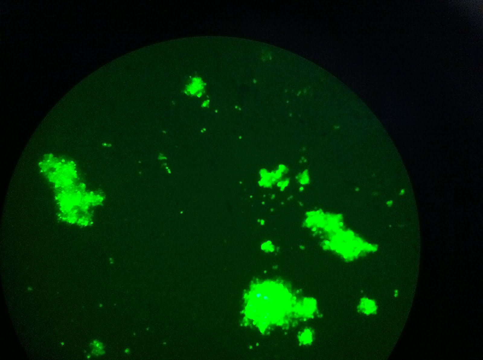
Below is the first round of pictures of cells expressing GFP. These pictures came from a single colony on an agar plate, which means I didn't transfer it to a liquid culture (i.e. it was sacrificed for science). However it does give us promise that we will have GFP-expressing lines of diatoms in the near future/ I've been working on growing up the cells in liquid cultures and hope to have something by the end of the week. After that, it's a matter of growing up larger liquid cultures before we can test some science!

The view through the microscope. So much green!

This frame shows some of the variability in the intensities of fluorescence. Denser cell populations will appear brighter.

Some clumps of cells are larger than others.

Saturday, August 20, 2011

DUN DUN DUN: DIATOM TRANSFORMATION (video edition)


I TRANSFORMED MY DIATOMS. ALL IS WELL. THIS IS AWESOME.

Well of course that is if we successfully transformed the diatoms. There's a 1-2 week grow period during which we have to wait in order to see transformed clones. I'll keep this blog posted and will post a text post regarding the transformation soon as well.