Saturday, September 17, 2011

The Genetic Transformation of Diatoms with Inducible Expression Plasmids

Last month my undergraduate/now graduate research project at Clark University reached a new level of awesome with the transformation of diatom cells with plasmids I've been working on for several semesters. It's been a long time coming for sure, with both problems encountered in the lab project and juggling other college courses.

In my blog I've chronicled some steps along the way on my project, and I've talked about:
And now I can talk about the next step of my project: the transformation of diatoms with the plasmids I've created.

Now I am undergoing the process of selecting lines of transformed diatoms for the next step in our project. But we've recently realized that the light intensity in our growth chamber is much brighter than previously published experiments growing transformed cultures of diatoms. Increasing light intensity reduces the amount of chlorophyll made by plants and algae. Why is this important? Two reasons:
  1. Less chlorophyll will reduce the pigmentation of our cells, making them harder to see once we transfer them to liquid culture.
  2. Our gene giving resistance to our antibiotic (that allows us to select for positive transformants) is driven by a chlorophyll-associated protein, which means expression may be reduced in higher light intensities. If chlorophyll expression is reduced by high light levels, then this may reduce the activity of the chlorophyll-associated protein that drives the antibiotic resistance in our diatoms, which means less resistance to the antibiotic in the media. This ultimately means reduced or no growth.
Now, our diatom cultures have been growing okay on their agar plates, but I'm beginning to wonder if they're growing slower in liquid cultures, where they're more likely to absorb more light. To combat this, I'm growing some liquid cultures behind layers of porch screen to reduce light levels penetrating the liquid cultures. In the below picture, you can see the different diatom cultures I have growing right now:

Transformed diatoms grow on agar plates (left foreground) an in liquid media (left background and right).
I've been growing diatoms in 3 mL cultures in little dishes, which you can see in the back left. Groups of 3 dishes sit in a petri dish to allow easier transportation. These dishes however require 3 mL of culture just to cover the entire bottom of the dish, which means the cells are going to be fairly diluted. I also started 1 mL cultures in the test tubes on the right, where porch screen blocks out a lot of the incoming light. Some of the 3 mL cultures also have screen on top of them to block out some of the light.

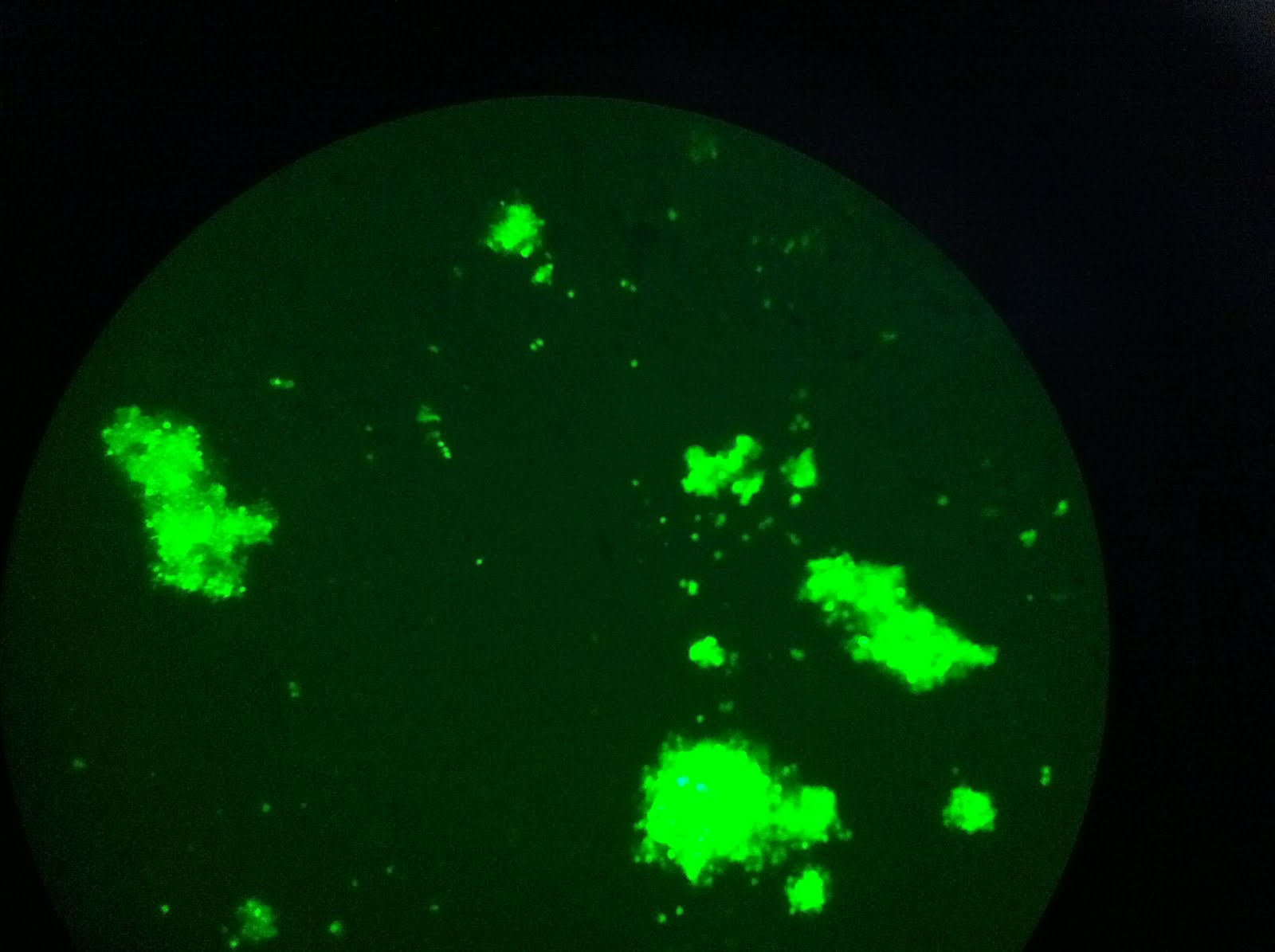
Aside from trying to grow our cells in liquid culture, I've been looking at them under a fluorescent microscope to try to determine whether our cells are fluorescing due to GFP expression or whether it's residual glow from chlorophyll. UV light from the microscope excites all pigments in the sample, and then I look at the sample through different filters that only allow certain wavelengths of light to show through. However, our current filter set up hasn't allowed a clear delineation between GFP and chlorophyll yet. Most of our pictures look something like this:

...but they usually have a lot more background color in there too.

My next steps will be to either design or order commercial GFP GSPs--gene specific primers for GFP. That way we can run a PCR on some diatom DNA and determine whether they have the GFP gene they are supposed to be transformed with. Because I have a great control (the original plasmid DNA), I can test a whole bunch of diatom colonies at once and be able to select appropriate lines of diatoms faster.

Tuesday, September 13, 2011

Glowing Green Cells

Just before the weekend, my professor wanted to look under the microscope to see whether our transformed diatom cells were expressing our reporter gene GFP. I've been culturing several different lines of transformed diatoms since the transformation. These cells were co-transformed, which means the resistance plasmid is separate from the GFP plasmid--that is the transformation process shot two different plasmids at the diatom cultures. Therefore we need to select for diatom lines that posses both plasmids: the resistance plasmid and the GFP plasmid.

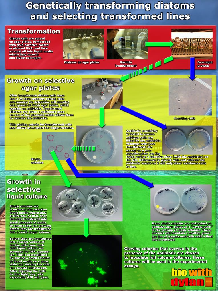
Below is a poster I made briefly describing each of the three main steps in the process: transformation, growing the diatoms on selective agar plates, and subculturing diatom cells in selective liquid cultures.

The diatoms will only grow if they received a copy of the resistance plasmid. And well, we can tell that the diatoms don't particularly like the antibiotic we put in the growth media:

By performing a simple antibiotic sensitivity test, we can show that diatoms will not survive in the presence of the antibiotic unless they receive the plasmid that confers resistance.

Below is the first round of pictures of cells expressing GFP. These pictures came from a single colony on an agar plate, which means I didn't transfer it to a liquid culture (i.e. it was sacrificed for science). However it does give us promise that we will have GFP-expressing lines of diatoms in the near future/ I've been working on growing up the cells in liquid cultures and hope to have something by the end of the week. After that, it's a matter of growing up larger liquid cultures before we can test some science!

The view through the microscope. So much green!

This frame shows some of the variability in the intensities of fluorescence. Denser cell populations will appear brighter.

Some clumps of cells are larger than others.

Sunday, September 4, 2011

The start of the semester & culturing transformed diatoms

The backside of Goddard Library, one of my favorite photo spots.
The Fall semester has quickly begun at Clark University. While I was worried that Hurricane Irene was going to make move in day for the sophomores, juniors, and seniors messy, Irene barely inflicted any serious weather upon Worcester (we were lucky). I've been working at the bookstore the past two weeks where it has been very busy, as students have been scrambling to get all of their books on time. As a result, I kind of missed the first week of craziness on campus for better or for worse. But I always love the start of the semester--everyone is always so happy to see each other and be back on campus.

On Friday, we had our annual student activities fair, where almost every social club on campus from CUFS (Clark U Film Society, which shows movies on Tuesdays and Thursdays in our cinema) to ROCU (Radio of Clark U) is represented by student members. This is a great way for incoming first years to discover all of what Clark has to offer for our ever expanding extracurricular groups.

A panorama of the student activities fair, as seen from Main St.
The fair from the window of Tilton Hall, nearly showing the fair in its entirety.
Additionally, I posted a new video on YouTube chronicling the start of the semester, Hurricane Irene in Worcester, and last but not least, my most recent lab work:


Thursday, September 1, 2011

Everybody's working for the weekend (or just grad school application money)

Recently, I've pretty much only had time for two things: working at the Clark bookstore and looking into grad schools. Because the bookstore is crazy busy the first few weeks or so, I've been working more than twice as much as I'd like to in order to help out. I sincerely hope things start to slow down really soon, because I've been working a lot the past two weeks. While this means I'll have money (which I can then spend on grad school applications), I've had little time to do anything else.

Coincidentally however, this is a perfect time to be busy at the bookstore when it comes to lab work because I'm in the process of growing my transformed diatoms on selective plates, a process that is going to take me at least two weeks before the next step. This next step may come as soon as tomorrow, before another one to two week growth period before I can even start to test my diatoms. But this isn't the topic of this post.

In my spare time when I'm not feeling completely exhausted from my tiring shifts at the bookstore, I've been looking at Ph.D. programs to start after I finish my Masters project here at Clark. This is actually a much scarier topic than I've lead myself to believe, because it doesn't take into account a lot of unknowns, such as whether I'll be able to get into grad school, whether I'll be able to finish my Masters project on time, and whether my girlfriend and I can get into grad schools remotely close to one another. While a lot of this is out of my control at this point and time, it doesn't make me feel better when I think about it. The best I can do at my point is get a first round of applications done as soon as possible so I can apply to my top schools as soon as possible. Applying to Ph.D. programs early is important, because there is only so much money at each school to support students. While I have to work at the bookstore again this weekend for an afternoon and I have some other things to clean up, I hope to have my act together on my first round of schools by early next week, and have contacted my the professors I'd like to work with the most.

It's embarrassing to admit, but I haven't even taken the GREs yet and I won't take them until mid-November, only a few weeks until some applications are due. Depending on how my applications are, I may have to bite the bullet and pay the stupid fee and bump up my exam day. Originally I was worried I was going to be weighed down with applications and lab work too much in order to properly study for the GREs. While that still may be the case, I'm wondering whether taking the GREs earlier would be better, so I can have a complete package application sooner.

That reminds me... I need to contact two more professors about writing me recommendations. Oh boy. This is what happens when my schedule becomes abnormal for two weeks thanks to working at a "real" job--I lose track of things. Along the same vein, it's weird how I feel like I've missed the start of school because I've been stuck at the bookstore the entire first week of school. *sigh* I've even started to panic that I'll miss the best weeks of fall, but I'll make sure that doesn't happen. I'm really hoping the bookstore will start to slow down really soon (as early as the middle of next week), so I can work less and get my act together in lab when the need arises.

Sunday, August 28, 2011

Lab Photos

I've been taking a lot of photographs in lab to post on Tumblr, as a quick way to share my work in the lab. Here is a recent collection of my favorites.

My bacteria have a habit of spelling out scientific messages, but this time they took it to a new level!

A plasmid DNA resuspension. Black permanent marker outlines the pelleted DNA from an ethanol precipitation at the bottom of three tubes, a trick I’ve started to use in order to aid the resuspension of DNA into a smaller, more concentrated volume. The outline allows me to precisely add a very small amount of buffer to the pelleted DNA, as it sits taped at an angle to a pipet tip box.

After completing a set of inducible expression plasmids which I transformed cultures of diatoms with this past week, it was time to throw out several months worth of agar plates. These agar plates were home to different lines of E. coli which I used to clone different parts of my plasmids. Now that I have glycerol stocks of these E. coli lines (frozen stocks of bacteria cultures stored at -80°C), I was able to clean out my bin of agar plates. Not only did it feel good to get rid of so many plates, but this is an exciting stage in my project as it starts in a new direction.

Genetically transformed diatoms scatter the surface of a selective agar plate. The agar plate, specially formulated to support the growth of the microscopic algae, is made with the antibiotic ClonNAT which is strong enough to prevent the growth of diatoms without a resistance plasmid. The genetic transformation was a co-transformation of a pair of plasmids: the inducible expression plasmid which we will use for genetic regulation tests, and a resistance plasmid for ClonNAT resistance.

This is a new “invention” I came up with the other day in lab: a simple pen/marker holder. While it’s only big enough for 2-3 markers, it’s perfect for our lab benches because our permanent markers wander off. This way our markers stay put on one side of the lab bench. I used clean (but previously used) 50mL conical tubes with a bit of lab tape.

Saturday, August 20, 2011

DUN DUN DUN: DIATOM TRANSFORMATION (video edition)


I TRANSFORMED MY DIATOMS. ALL IS WELL. THIS IS AWESOME.

Well of course that is if we successfully transformed the diatoms. There's a 1-2 week grow period during which we have to wait in order to see transformed clones. I'll keep this blog posted and will post a text post regarding the transformation soon as well.

Tumblr & Science

I've created a Tumblr page to post cool pictures from lab to run along side my blog here about my project and you should really check it out:

This is where I post stuff like this that I've made:

But really, I should be posting cool stuff like that here as well, which I will do in the future.

Sunday, August 14, 2011

Building inducible expression plasmids (aka Science)

Below is a video I've been trying to publish for a long time coming this summer, but because my summer project only came to a close this week, I've been hesitant to post anything.


In this video, I give an update on my lab work and discuss my overall lab project, which is a great review for myself and my viewers. This is an exciting time for me at Clark University, because I’m beginning the real stages of becoming a graduate student in our accelerated Master’s program. After conducting undergraduate research, I’m using it to support a 5th year of schooling at Clark to finish up my Master’s.

My project concerns the 3’ UTR of the nitrogen-assimilating genes nitrate reductase and nitrite reductase in diatoms (if you haven't read up on my blog before), because we have evidence that along this region these genes are up- and down-regulated by environmental conditions. To test this hypothesis, I’ve created plasmids that drive expression of a reporter gene (GFP) with promoter and terminator regions derived from diatom genes. We’ll compare the activity of GFP with differing 3’ UTR elements to determine whether this region of the nitrogen-assimilating genes is crucial in their regulation.

For the gene nitrate reductase, this is old news. But it is news for my blog that I've completed the plasmids for nitrite reductase, which is really the story of my video.

I'm really excited to be where I am in my project. This week, I hope to genetically transform my diatoms with the plasmids I've created. Oh baby Darwin, this could be a HUGE week for me and my scientific career! I'll be back soon with transformation news.

Thursday, August 11, 2011

The completion of my NiR plasmids

This summer's project has been to complete inducible expression plasmids to test the regulation of nitrite reductase (NiR) in the diatom Thalassiosira pseudonana. Inducible expression means we can induce the expression of a gene located on a plasmid based on environmental factors. These plasmids will be used to test whether the 3' end of the NiR plays a role in regulation the gene's activity based on environmental cues. We'll compare the activity of our reporter gene (GFP) from our control plasmid (with an NiR terminating region) and our developing experimental plasmid (with an Actin terminating region, as actin has no role with nitrogen assimilation).

An analytical double digest and gel to determine which of my bacteria colonies actually had the plasmid I wanted. My colony screen gave me some funny results, so I wanted to double check with a digest. The digest confirmed several of my colonies appear to have the plasmid and correct insert, giving me the green light to grow up more bacteria cultures and prep and purify their plasmids.

While it appears my NiR-NiR plasmid is completed (sections of the NiR gene flank the reporter gene in the plasmid), the completion of the experimental plasmid is right behind the control plasmid. Today I transformed some bacteria with what I hope is the complete experimental plasmid. I should know by tomorrow afternoon if I have what I think I do!

Meanwhile, I have scheduled a date at the University of Rhode Island to transform my diatoms! While I may only transform the diatoms with my set of nitrate reductase (NR, a different but related gene) plasmids (both with an NR terminus and an actin terminus, like the NiR plasmids I'm finishing up now).

Wednesday, August 3, 2011

This DNA has a sequence and I have every intention to find out what it is! (and other stories)

In my quest to amplify a region of the nitrite reductase (NiR) gene that I can then ligate into a plasmid for in vivo testing, I needed to sequence my portion of DNA for three main reasons:
  1. I needed to make sure that the restriction sites I added with gene specific primers were added to either end of the sequence. These restriction sites will allow me to cut out my amplified piece of DNA that is cloned into a cloning vector.
  2. I need to verify that the sequence of DNA I've been cloning is indeed from the NiR gene, especially since the DNA sequence has been marginally longer on my agarose gels than anticipated.
  3. Previous data acquired from a database told me I should be able to get a restriction enzyme to cut somewhere in the middle of the DNA sequence I had cloned, but the restriction enzyme wouldn't cut in the middle (but it did cut on either side of the DNA sequence that I cloned into a vector, as the vector had that restriction site on either side of the multiple cloning site). Essentially, that restriction site didn't exist in my DNA sequence like it should have.
So, couple weeks ago, I performed a PCR cycle sequencing reaction to start the process of sequencing. The purpose of this sequencing reaction is to amplify a region of DNA with DNA bases that have special tags. These tags are read by a sequencing machine yielding a series of base signals like below:

The color coded lines refer to a single base category (either A, T, G or C) and denote the signal strength at that spot in the sequence. Based on the predominant signal at a spot in the sequence, the software makes a judgment call as to which base is present at that location. Often it is quite clear to the machine and software that a particular letter of DNA is certainly an A at a specific location because there is a single strong signal peak. Other times there may be several signal peaks at a single location because of a lower quality sequencing reaction (e.g. there is too much or too little DNA to be read properly), which make it harder to determine which DNA letter actually exists in the DNA sequence.

In order for the DNA to be read by the sequencing machine, it needs to be single stranded. This means we make up separate PCR reactions with only one primer, which means that the one primer in a PCR reaction will attach to the DNA and copy the DNA until it falls off. To make it easier on myself, I followed a standard protocol that sequences a section of DNA that has been cloned into a plasmid vector. This way there is a buffer of vector plasmid sequence on either side of my DNA sequence of interest, where the primers that pair with the vector plasmid would bind.

I performed my PCR reaction and prepared it for the sequencing machine and was about to load my DNA onto the DNA sequencing plate (which goes into the machine), when I was notified that the machine was down and I couldn't sequence that day or possibly for the rest of the week. It took me a week or more for my samples to get read, but they finally did get read and my sequences were good enough to give me some answers.

I use a special computer program that will align the DNA sequences to one another. Running multiple samples of the same sequence will allow the program to compare answers and yield a single, best result sequence. Interestingly enough, while my four samples all matched each other, there was a small region (about 20 bases) that did not match up with the database sequence for my gene. This may have been why my restriction site wouldn't cut--because it wasn't there. One of my labmates is going to sequence the same gene soon, so I will be able to compare my results with hers and determine whether the predicted sequence in the database needs to be updated.

I can then take the sequence from the first program (the results) and pop it into another program, to quickly find all of the restriction sites that are present.
Here I can see that my BamHI and NotI restriction sites are indeed at either end of my sequence, with the EcoRI sites just outside of the Bam and Not sites. Because the EcoRI sites are in the vector which I cloned my sequence with, I know that the Bam and Not sites are on either side of my sequence like they should be.

At present I have ligated this section of DNA (the piece of the NiR gene) with the other portion of the NiR plasmid, and will transform some bacteria today. If the plasmid worked and was successfully cloned into my cells, they bacteria will be able to grow on the plate laced with ampicillin. Here's hoping!